Peritoneal And Subcutaneous Hemorrhage
The dura mater and the lock membrane () are two of the three human meninges. The meninges are structures that contain the central nervous system. Subdural hematoma, or subarachnoid hemorrhage, or subarachnoid hemorrhage (SAV), refers to hemorrhage that occurs below these two meninges.
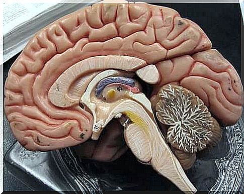
Meninges
The brain and spinal cord are vital parts of the body protected by the skull and spine, but there is also another protection mechanism around them: the meninges, which are also involved in nerve development.
As the US Social Affairs and the Ministry of Health NIH Cancer Dictionary (Dictionary of Cancer ) to determine the ” meninges consist of three layers of tissue that cover and protect the brain and spinal cord. “ In other words, there are three different meninges in our brain, which are named from the outside in the following order:
- Hard membrane or dura ( Dura mater)
- Locking membrane or arachnoid membrane (A rachnoidea)
- Soft film or soft film ()
The most outermost and thickest of these meninges is the dura, or dura, which is separated from the bone by the epidural space. In the skull, this state is so-called virtual, that is, it does not exist, because the sclera is attached to the skull. However, the epidural space is found in the spinal cord and is filled with blood vessels and fat.
Below the dura, there is another meninges, the locking membrane, which is separated from the bone by the subdural space. This state is also virtual and only becomes real when there is bleeding in the brain that separates the sclera and the sclera.
The lock membrane sends a series of extensions to the soft membrane that pass through the lock membrane cavity, i.e., the subarachnoid space. The subarachnoid space is filled with cerebrospinal fluid and its function is, among other things, to soften and dampen pressure changes caused by shocks or sudden movements.
Finally, the soft film is closely attached to the nerve tissue and follows it tight even in its smallest and narrowest lines and grooves. The possibility of the soft film extending even into the interior of the tissue is also currently being investigated.
Peritoneal and subcutaneous hemorrhage
Pericardial hemorrhage and interlocking hemorrhage occur when blood flowing in blood vessels leaks out and is stored between different meninges, as the case may be. This in turn damages the brain tissue, leading to various clinical cases.
Depending on whether the bleeding occurs in the subchondral state or in the subchondral state, the factors that trigger the bleeding, the course of the pathology, and its diagnosis differ.
Peritoneal haemorrhage or subdural hematoma
Subarachnoid hemorrhage is defined as hemorrhage that accumulates in the virtual space between the sclera and the sclera. This blood usually comes from blood vessels that have responded to a head attack or other injury. However, subcutaneous hemorrhage can be found in three different types depending on the time it takes for the hemorrhage to develop.
- Acute subcutaneous bleeding
- Subacute duodenal bleeding
- Chronic subcutaneous hemorrhage
Acute subcutaneous bleeding
Acute subarachnoid hemorrhage gives signs of itself at an early stage. It is usually a severe head injury that breaks the blood vessels that flow from the cortex to the meninges.
A patient with acute subcutaneous hemorrhage usually falls immediately into a coma. In addition, he usually shows signs associated with focal hemisphere. In other words, this means that a certain part of the brain stops working. Some signs of cerebral focal focus include:
- Hemiparesis, or partial paralysis: A partial inability to move a specific part of the body due to an injury to the area responsible for motor skills.
- Mydriasis: An abnormal enlargement of the pupil diameter due to an injury to the area that controls the iris muscles.
Subacute duodenal bleeding
The development of subacute duodenal bleeding is somewhat slower and usually less severe than acute bleeding. This is because there is usually less bleeding, so that the mechanisms that promote blood clotting can even stop the bleeding completely. The cause of subacute subarachnoid hemorrhage is also usually due to injury.
In subacute duodenal bleeding, the patient often loses consciousness but recovers later. Over the next few days, he begins to experience a gradual increase in blurred vision as well as other signs of neurological focality.
Chronic subcutaneous hemorrhage
Chronic subcutaneous hemorrhage is the result of several minor injuries that have accumulated over time. They cause small blood extravasations and, when not reabsorbed into the blood vessels, lead to a significant scale of subcutaneous hemorrhage. Chronic duodenal bleeding is relatively common in the elderly.
An early symptom of chronic duodenal hemorrhage is usually a headache associated with changes in stimulus response and behavior. In addition, brain function gradually deteriorates, making a person often tired and sleepy, and thinking slows down among many other symptoms.
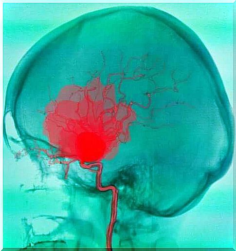
Subarachnoid hemorrhage or subarachnoid hemorrhage (SAV)
In subcutaneous hemorrhage, blood collects in the space between the diaphragm and the soft membrane. The blood usually comes from the arteries and the bleeding can be caused by a number of different reasons. The most common cause of submucosal hemorrhage is a rupture of the aneurysm, or artery bulge, but in some cases the hemorrhage may also be due to a deformity of the blood vessel.
Aneurysms can manifest as headaches or epileptic seizures before they rupture. In as many as a third of aneurysm-related cases, the factor that led to rupture is physical exertion with an emotional component or prolonged exposure to the sun.

When the aneurysm ruptures, subarachnoid hemorrhage begins. This usually happens in adults aged 40-60 years. The onset of bleeding is sudden and the patient develops the following symptoms as a result of the bleeding:
- The patient vomits
- The patient experiences visual disturbances and blurred vision
- The patient experiences a very severe headache
- Patient suffers from photophobia (eyes do not tolerate light due to pain or other discomfort caused by light)
Approximately 48 hours after the onset of bleeding, the patient may in many cases also have meningeal syndrome due to bleeding-induced irritation of the meninges. In this case, in addition to the above symptoms, the patient also suffers from neck stiffness. In addition, the patient may experience neurological disorders in a particular part of the body, such as paralysis of eye movements.
Subarachnoid hemorrhage causes sequelae in up to 60 percent of patients. In addition, 40 percent of survivors develop some form of addiction.